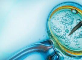
PREIMPLANTATION DIAGNOSIS (PGD)
PGD (Preimplantation Genetic Diagnosis) for single gene disorders is a genetic test that may be performed during assisted reproduction treatment to screen embryos that are at risk to develop a serious genetic disorder.
PGD is performed on a small embryo biopsy and identifies which embryos are not at increased risk of developing the disease.
Some genetic diseases only affect one sex rather than the other. In this case, the embryo is tested to find out its sex and only embryos of the non-affected sex are transferred to the mother’s womb.
PGD is recommended for:
- couples at risk of transmitting chromosomal alterations or monogenic diseases,
- couples with a medical history of repeated miscarriages,
- couples with several cycles of IVF that have not achieved pregnancy,
- when are abnormalities in spermatozoa maturation and
- women of advanced age.
PGD Procedure
The main steps in an IVF/PGD cycle include:
- Fertility drugs are administered to a woman
These may be given orally, or by injection over a 2 to 4-week period. The fertility drugs act to stimulate more than one ovarian follicle to mature and provide a number of eggs for fertilisation.
- Blood and ultrasound tests
These are used to check the woman’s follicle development so that egg collection can be timed. These tests are also to avoid overstimulating the ovaries.
- Egg collection
The woman is placed under a light anaesthetic or sedation (and occasionally a general anaesthetic) so that eggs may be collected with the help of a vaginal ultrasound probe. This will be performed by a gynaecologist.
- Fertilization
After egg collection, the eggs will be fertilised with sperm in the laboratory through IVF or ICSI. Sperm may have been freshly collected or previously collected and frozen. The fertilised eggs are matured or ‘cultured’ until 3 or 5 days of age.
- Biopsy (removal of cells for testing)
Cell removal is carried out on embryos at 3 days of age when an embryo will usually have between 6 to 8 cells.
A laser is used to open the outside shell of the embryo, and the cell/s are carefully separated from the remaining embryonic cells. In most cases, the ‘hole’ or deficit will close over, and the embryo
will develop as it should.
However, a very small percentage of embryos will not continue to develop following embryo biopsy.
- Laboratory analysis
The biopsied cells are then prepared and tested for the specific gene or translocation.
Polymerase chain reaction (PCR) is the most common laboratory method used for PGD.
When embryos are tested on day 3, it may be possible to transfer the embryos after biopsy/PGD as the PGD results are usually back within 2 days. When day 5 (blastocyst) embryos are tested, they will require freezing following the biopsy in order to await results.
- Transfer
If the PGD results show that an embryo is suitable for transfer, the embryo are transferred using a catheter that has been guided into the woman’s uterus. Any remaining embryos that are suitable for transfer can be frozen and stored for later use.


